Lead Biomedical Scientist and British Association for Cytopathology Executive Committee Member Hedley Glencross introduces a project to improve the outdated cervical screening programme in the Republic of Moldova.

For nearly three years I have been involved with a small project team concerned with introducing an organised cervical screening programme to the Republic of Moldova (RM). This has been an interesting project, not only coming at a time when UK cervical screening programmes are stopping cytology-based screening in the midst of a move to primary HPV testing, but also encountering the obvious cultural differences between the UK and RM.
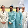
I have visited RM three times, conducting laboratory assessments and participating in a wider cervical cytology education and training programme, delivering lectures and workshops.

RM is a landlocked eastern European country that is surrounded by Ukraine and Romania. It is a poor country – one of the poorest in Europe – and has a mostly rural-based economy. The most recent figures show RM had a population estimated at just under three million in 2016. Out of the main urban areas, water tends to be drawn from wells and there is little or no electricity or mains sanitation.
Cervical cancer is a large burden in the country, with approximately three women, mostly younger women, a week dying from cervical cancer. Although cervical cytology does exist in RM, and it has been claimed that in 2015, 250,000 smears were “screened”, enough to result in a 70% coverage rate of the population, based on a three-yearly screening programme, the incidence of and mortality rates from cervical cancer remain stubbornly high.
This disparity may be explained by a number of factors, not least the distinct lack of training in the screening and interpretation of cervical smears, but also the way in which smears are taken and stained. Almost contemporaneously with Papanicolaou, a Romanian pathologist, Aurel Babes published a paper on the diagnosis of cervical cancer using air-dried smears and Romanowsky/Giemsa (R/G) staining. Unfortunately, Babes published this paper in a French language journal and did no further work on this matter, so went largely unnoticed in English-speaking and wider European countries. His work though persisted in eastern Europe, especially during the cold war when much of eastern Europe was in effect closed to outside influences.
While R/G staining has a valuable place in diagnostic cytology, the often subtle chromatin disturbances, cellular variation and active keratinisation seen in dyskaryosis are largely “invisible”, making accurate identification of CIN 3 very difficult. Left untreated CIN 3 will progress to cervical cancer in approximately a third of women after 10 years. I did see Papanicolaou-stained smears as well, but their quality left a lot to be desired.
Screening programme
The laboratory facilities were rudimentary at best and although there was some (very) modern equipment seen, it was either underused or not used at all. Coupled with this observation was also the lack
of quality measures we take for granted at both the smear-taking and the screening and reporting stages. Fortunately, the project group was able to call on the expertise of UK-based cytopathologists and histopathologists, as well as myself and a number of committed and enthusiastic Moldovan-based gynaecologists and pathologists, all led and driven by the International Cervical Cancer Prevention Association (ICCPA).
As RM is a resource-limited country, a decision was made to base, at least initially, this screening programme on conventional cytology smears taken using a Cervex-Brush and reported using modified Bethesda terminology. Although this may seem an odd decision, given international moves to primary HPV testing and vaccination, historical data from the UK and other countries have shown the large positive impact on cervical cancer incidence and mortality rates following the introduction of such screening programmes. What the project team has been able to achieve includes:
- Training and converting primary health clinics to accurately take and fix cervical smears, ensuring each woman is uniquely identified and smears are properly labelled
- Giving screening laboratories a working Papanicolaou staining method that allows proper visualisation of smears
- Delivering a number of training courses and workshops in cytology screening and reporting
- Delivering a number of courses and workshops in histopathology reporting.
While there is much work to do regarding computerisation, call/recall, fail-safe, quality assurance and many other aspects of the programme, the work done so far is a sound base upon which to build.
One of the next planned phases for the laboratory part of the project, alongside further cytology training, will be to address histological laboratory matters, including fixation, block selection, tissue processing, embedding, cutting and staining procedures. Work on setting up this part of the project is on-going, which will likely involve reciprocal visits between UK and RM to train one of their staff, who will then cascade this to other laboratories in RM.
This has been and continues to be an interesting experience for me, not only working outside the UK, but also my first encounter with simultaneous translation of lectures. I have met and worked with a brilliant group of people. Special thanks must go to Dr Philip Davies (ICCPA), Drs Mary Brett and Mike Coutts (UK), Drs Eugen Melnic, Ruslan Pretula and Ecaterina Foca (RM), plus all of the medical and laboratory staff from RM, who are too many to name individually, but who have been a wonderful group of students. Personally, I hope to continue with this project and help see it through to a successful outcome.
Hedley Glencross is Lead Biomedical Scientist in Diagnostic Cytology and Andrology at Queen Alexandra Hospital, Portsmouth. He is an Executive Committee member of the British Association for Cytopathology.
Image credit | Getty | Shutterstock