Scientists have developed a new imaging method to see where antibiotics have reached bacteria within tissues.
It could be used to help develop more effective antibiotic treatments, reducing the risk of antibiotic resistance.
During bacterial infections, bacteria enter human cells, which poses a challenge for treatment.
If researchers could select for or develop more effective antibiotics based on where they reach, this may reduce the length of treatment needed, which in turn could reduce the risk of antibiotic resistance developing.
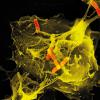
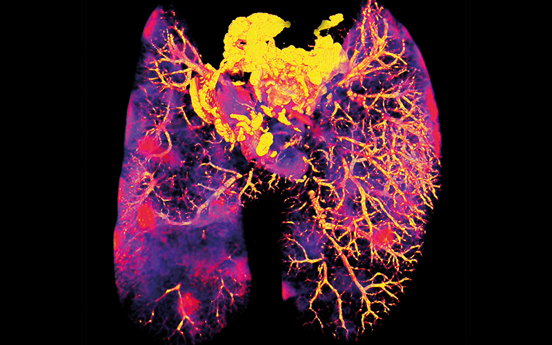
To develop the imaging method the researcher analysed lung tissue from mice with tuberculosis and treated with the antibiotic bedaquiline.
They combined a variety of imaging methods, including confocal laser scanning microscopy, 3D fluorescence microscopy, electron microscopy and nanoscale secondary ion mass spectrometry, to develop their new approach. They found that bedaquiline had not reached all infected cells in the lung tissue and also had not entered all infected areas within infected cells.
They also found this antibiotic was collecting in macrophages and in polymorphonuclear cells, both types of immune cell. This was a surprise as these cells have different environments and it wasn’t thought that one antibiotic would be able to enter both.